B-Scan Ultrasonography
|
Troia Eye & Laser PC is utilizing the DGH Technology, Inc. Scanmate B-Scan (DGH8000) the most advanced ultrasound technology available today.
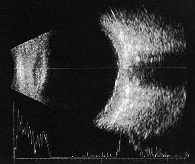
B-Scan ultrasonography (Ocular Ultrasound), is a diagnostic test used in modern optometry to produce a two-dimensional, cross-sectional view of the eye and the orbit.
B-Scan ultrasonography is wheelchair accessible and an important adjuvant for the clinical assessment of a variety of ocular and orbital diseases. B-Scan ultrasonography testing can gather a vast amount of information not possible with clinical examination alone. This instrumentation is designed to provide a general understanding of echographic characteristics of various ocular pathologies.
|
Ultrasound, also referred to as echography, uses high frequency sound waves to produce images of the internal eye structures. It is a helpful diagnostic tool if cataracts or other conditions prevent a doctor from viewing inside of your eye with traditional methods. Ultrasound is helpful for diagnosing retinal detachment, vitreous bleeding, tumors, inflammation, lesions in the eye socket bone, or foreign bodies in the eye.
Ultrasound is a quick and painless procedure. A small transducer device is placed on your eye. The transducer transmits sound waves to a computer that produces images of the structures inside your eye. The B-scan is done through the closed eyelids and requires no preparation. You will be instructed to look in various directions during the B-scan ultrasound. This allows Vincent Troia, O.D. to view your inner eye from different angles. The procedure is performed at Troia Eye & Laser P.C., and allows Vincent Troia, O.D. to evaluate your results immediately during your comprehensive ocular exam.